INTRODUCTION
Thymoma and thymic carcinoma is a rare tumor originating from epithelial cells of the thymus. Metastasis occurs mainly in the local site or thoracic cavity2). Spine metastasis was reported in 7 cases3,5,6,8,10,12). These cases were easily distinguished from metastasis by destroying the surrounding vertebral bone. However, our case was uniquely limited to the epidural space and invaded the intervertebral foramen.
CASE REPORT
A 78-year-old man developed weakness of the left wrist and grasp (grade 2). He experienced pain for 2 days, but there was no pain at admission. The patient was diagnosed with malignant thymic tumor with involvement of the lungs and underwent surgical resection 7 years ago. Histopathologically, malignant thymoma was diagnosed with combined thyroid B2 and B3. Therefore, additional radiotherapy was performed. The tumor did not recur and the patient was cured.
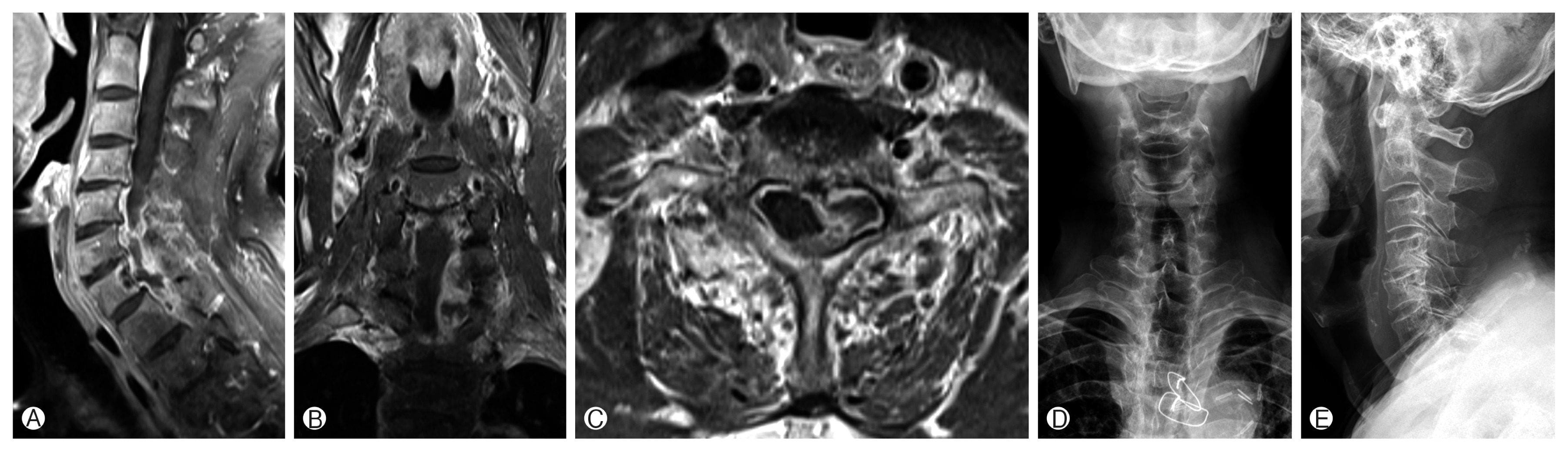
Cervical spine magnetic resonance imaging (MRI) revealed a rim-enhanced lesion compressing the cord into the C6–7–T1 epidural space and invading the C7–T1 intervertebral for a-men. MRI with contrast showed low signal lesions in the vertebral bone of C7 (Fig. 1). A laboratory study was performed at admission. White blood count, erythrocyte sedimentation rate, and C-reactive protein levels were normal.
A nearly total resection preserving nerve roots was performed via a total C6–C7–T1 laminectomy.
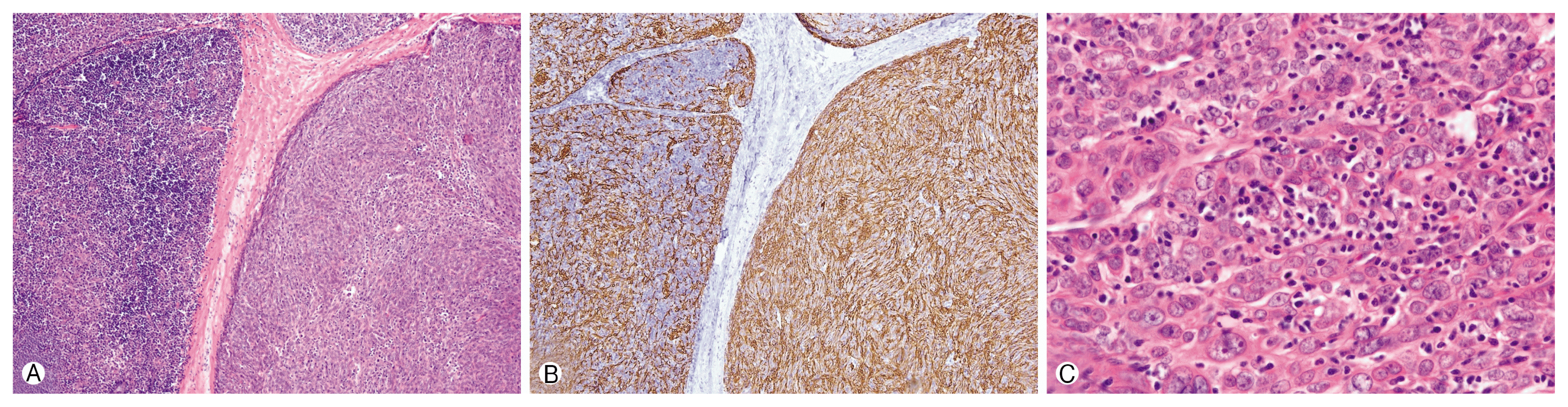
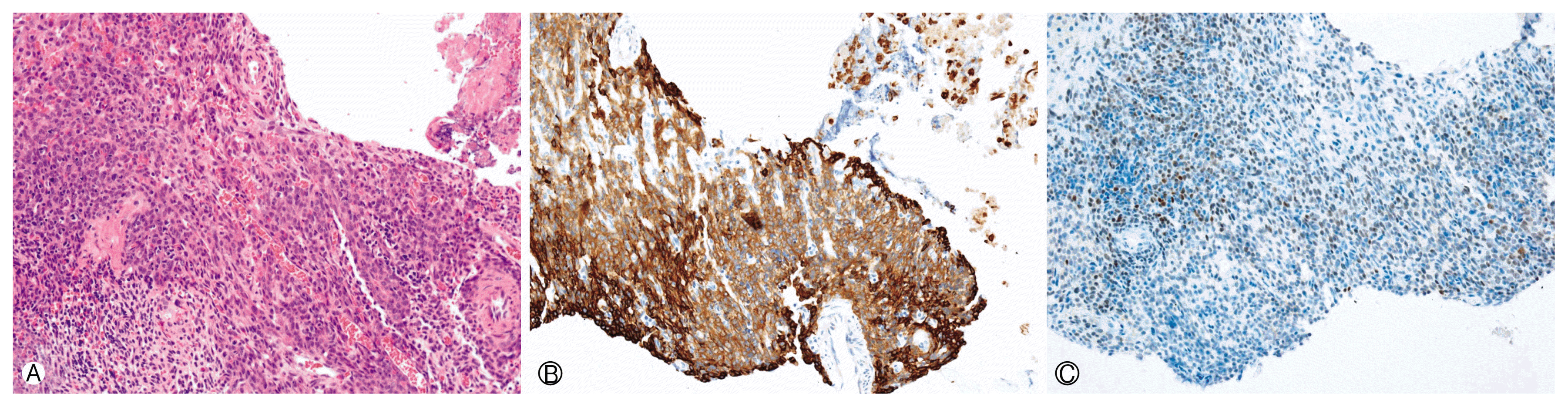
Histopathologically, the malignant thymoma that was operated on 7 years ago was composed of lobules separated by fibrous scar, and necrosis was observed in some of them. There were 2 types of tumor lobules: some lobules were mixed with lymphocytes of tumor epithelium, and some lobules were mostly tumor epithelial cells and lymphocytes were rarely observed (Fig. 2). The nuclei of tumor cells were round or ovoid, and the boundaries of cells were unclear. The nucleus was vesicular, indistinct, or distinct. Mitosis was rare. Tumor cells were positive for epithelial cell markers, including cytokeratin and epithelial membrane antigen, and negative for CD5 indicating B2 and B3 type thymoma. The tumor was invading the lungs and no lymph node metastasis was observed. Cervical lesions were accompanied by necrosis or bleeding. Tumor cells mixed with lymphocytes were observed. Tumor nuclei were round or ovoid and nonnodular (Fig. 3). Tumor cells were positive for epithelial cytokeratin and positive for PAX8, a thymic epithelial cell marker, and a patient was diagnosed with recurrent (metastatic) malignant thymoma.
We explained to the patient and patient’s family about the patient’s systemic condition and side effects of radiotherapy and chemotherapy. However, the patient and his family refused further treatment.
DISCUSSION
Thymoma and thymic carcinoma are uncommon epithelial lesions that originate from the thymus gland2). The incidence of thymomas has been estimated at 0.13 per 100,000 person in a year1). On the basis of the appearance of epithelial cells, the World Health Organization unified classification proposed 3 histological types of thymomas (types A, B, and C), and 5 classes (medullary, mixed, lymphocytic, cortical, and epithelial)13). Moran and Suster9) differentiated thymomas according to atypia of the neoplastic epithelial cells (type A–B2, well differentiated thymomas; type B3, atypical thymoma; type C, thymic carcinoma). Although thymic carcinomas are classified as type C in the World Health Organization classification, these tumors are not just another variant of thymoma.
In 6 cases reported previously, extradural lesions that were close to the spinal canal in MRI were compressing the dura mater and invading the paravertebral muscles3,5,6,8,10,12). In 1 case, intradural extramedullary metastasis was present after surgical treatment with extradural mass. After gadolinium administration, tumors showed strong enhancement. In computed tomography (CT) study, infiltrated vertebral bodies can show both as osteoplastic and osteolytic lesions. Our case was different from the case reported previously. MRI showed that the tumor was rim-enhanced, and CT showed that the tumor was not invading the vertebral body (Table 1).
Local spreading occurs rapidly in thymoma but distant metastasis occurs late. The distant spinal metastasis of thymoma requires an average of 11 years (4 to 17 years)8). Our case also developed distant metastasis after 7 years.
The 5-year survival rate of patients with distant metastasis of thymoma varied widely between 13.3% and 81% after multimodality treatment, including surgical resection of primary tumor, pleurectomy, chemotherapy, and irradiation14). However, surgical resection is the most important treatment for thymoma metastasis. In recurrent thymoma, reoperation is more effective at increasing the 5-year survival rate than radiation and chemotherapy7). The reoperation is aggressively recommended if it is possible to resect the lesion completely. Overall 5-year survival rates of the recurrence cases without reoperation were 36% and 51%, respectively, whereas the 5-year survival rates of the recurrence cases with reoperation were 47% and 64%, respectively. Also, overall 10-year survival rates of the recurrence cases without reoperation were 17 % and 43%, respectively, whereas the 10-year survival rates of the recurrence cases with reoperation were 35% and 53%, respectively4,11). In metastatic thymoma, surgical treatment is also more important than other treatments.
CONCLUSION
Spinal metastasis of thymoma is rare and occurs a few years later. The previous reported case occurred with involvement of the vertebral body or posterior element, but our case was purely rim-enhanced and appeared as an abscess and intradural extramedullary tumor. In addition, if there is a spinal epidural lesion, distant metastasis due to underlying disease should be considered.